Knee
Knee Anatomy
The knee is a complex joint made up of different structures including bones, tendons, ligaments and muscles. They all work together to maintain normal function and provide stability to the knee during movement.
Having a well-functioning healthy knee is essential for our mobility and ability to participate in various activities. Understanding the anatomy of the knee enhances your ability to discuss and choose the right treatment procedure for knee problems with your doctor.
Bones of the Knee
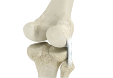
The knee is a hinge joint made up of two bones, the thighbone (femur) and the shinbone (tibia). There are two round knobs at the end of the femur called femoral condyles which articulate with the flat surface of the tibia called the tibial plateau. The tibia plateau on the inside of the leg is called the medial tibial plateau, and on the outside of the leg it is called the lateral tibial plateau.
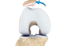
The two femoral condyles form a groove on the front (anterior) side of the knee called the patellofemoral groove. A small bone called the patella sits in this groove and forms the kneecap. It acts as a shield and protects the knee joint from direct trauma.
A fourth bone called the fibula is the other bone of the lower leg. This forms a small joint with the tibia. This joint has very little movement and is not considered a part of the main joint of the knee.
Articular Cartilage and Menisci of the Knee
Movement of the bones causes friction between the articulating surfaces. To reduce this friction, all articulating surfaces involved in movement are covered with a white, shiny, slippery layer called articular cartilage. The articulating surface of the femoral condyles, tibial plateaus and the back of the patella are covered with this cartilage. The cartilage provides a smooth surface that facilitates easy movement.
To further reduce friction between the articulating surfaces of the bones, the knee joint is lined by a synovial membrane which produces a thick clear fluid called synovial fluid. This fluid lubricates and nourishes the cartilage and bones inside the joint capsule.
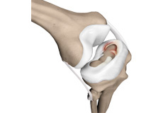
Within the knee joint, between the femur and tibia, there are two C shaped cartilaginous structures called menisci. Menisci function to provide stability to the knee by spreading the weight of the upper body across the whole surface of the tibial plateau. The menisci help in load- bearing by preventing the weight from concentrating onto a small area, which could damage the articular cartilage. The menisci also act as a cushion between the femur and tibia by absorbing the shock produced by activities such as walking, running and jumping.
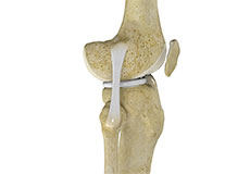
Ligaments of the Knee
Ligaments are tough bands of tissue that connect one bone to another bone. The ligaments of the knee function to stabilize the knee joint. There are two important groups of ligaments that hold the bones of the knee joint together, collateral ligaments and the cruciate ligament.
Collateral ligaments are present on either side of the knee. They function to prevent the knee from moving too far during side to side motion. The collateral ligament on the inside is called the medial collateral ligament (MCL) and the collateral ligament on the outside is called the lateral collateral ligament (LCL).
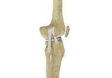
Cruciate ligaments, present inside the knee joint, control the back-and-forth motion of the knee. The cruciate ligament in the front of the knee is called anterior cruciate ligament or ACL and the cruciate ligament in the back of the knee is called posterior cruciate ligament or PCL.
Muscles of the Knee
Muscles: There are two major muscles, the quadriceps and the hamstrings, which enable movement of the knee joint. The quadriceps muscles are in the front of the thigh. When the quadriceps muscles contract, the knee straightens. The hamstrings are in the back of the thigh. When the hamstring muscles contract, the knee bends.
Tendons of the Knee
Tendons are structures that attach muscles to the bone. The quadriceps muscles of the knee meet just above the patella and attach to it through a tendon called the quadriceps tendon. The patella further attaches to the tibia through a tendon called the patella tendon. The quadriceps muscle, quadriceps tendon and patellar tendon all work together to straighten the knee. Similarly, the hamstring muscles at the back of the leg are attached to the knee joint with the hamstring tendon.

Meniscal Tears
Meniscus tear is the commonest knee injury in athletes, especially those involved in contact sports. A suddenly bend or twist in your knee cause the meniscus to tear. This is a traumatic meniscus tear. Elderly people are more prone to degenerative meniscal tears as the cartilage wears out and weakens with age.

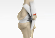
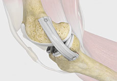
Patellar Tendon Rupture
Patellar tendon rupture is the rupture of the tendon that connects the patella (kneecap) to the top portion of the tibia (shinbone). The patellar tendon works together with the quadriceps muscle and the quadriceps tendon to allow your knee to straighten out.
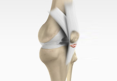
Quadriceps Tendon Rupture
Quadriceps tendon is a thick tissue located at the top of the kneecap. The quadriceps tendon works together with the quadriceps muscles to allow us to straighten our leg. The quadriceps muscles are the muscles located in front of the thigh.
Ligament Tears
The knee is a complex joint which consists of bone, cartilage, ligaments and tendons that make joint movements easy and at the same time more susceptible to various kinds of injuries.
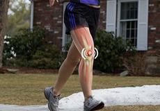
Runner’s Knee
Patellofemoral pain syndrome also called runner’s knee refers to pain under and around your kneecap. Patellofemoral pain is seen in number of medical conditions such as anterior knee pain syndrome, patellofemoral malalignment, and chondromalacia patella that cause pain around the front of the knee.
Knee Injury
Pain, swelling and stiffness are the common symptoms of any damage or injury to the knee. If care is not taken during the initial phases of injury, it may lead to joint damage that may end up destroying your knee.
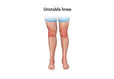
Unstable Knee
The knee joint is one of the largest joints in the body. This highly complex joint has several tissues supporting and stabilizing its movement:
Knee Sprain
Knee sprain is a common injury that occurs from overstretching of the ligaments that support the knee joint. A knee sprain occurs when the knee ligaments are twisted or turned beyond its normal range causing the ligaments to tear.
Patellar Instability
Patellar (knee cap) instability results from one or more dislocations or partial dislocations (subluxations). Patella is the small piece of bone in front of the knee that slides up and down the femoral groove (groove in the femur bone) during bending and stretching movements. The ligaments on the inner and outer sides of patella hold it in the femoral groove and avoid dislocation of patella from the groove.
Posterolateral Corner Injuries
Posterolateral corner injury is damage or injury to the structures of the posterolateral corner. The structures of the posterolateral corner include the lateral collateral ligament, the popliteus tendon, and the popliteo-fibular ligament. Injuries to the posterolateral corner most often occur with athletic trauma, motor-vehicle accidents and falls. An isolated injury to the posterolateral corner is rare, but often occurs with injuries to the cruciate ligaments, the anterior cruciate ligament (ACL) and the posterior cruciate ligament (PCL).
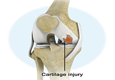
Cartilage Injuries
Articular or hyaline cartilage is the tissue lining the surface of the two bones in the knee joint. Cartilage helps the bones move smoothly against each other and can withstand the weight of the body during activities such as running and jumping. Articular cartilage does not have a direct blood supply to it so has little capacity to repair itself. Once the cartilage is torn it will not heal easily and can lead to degeneration of the articular surface, leading to development of osteoarthritis.
Non-surgical Treatments
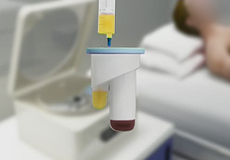
Platelet Rich Plasma/PRP
Our blood consists of a liquid component known as plasma. It also consists of three main solid components which include red blood cells (RBCs), white blood cells (WBCs), and platelets. Platelets play an important role in forming blood clots. They also consist of special proteins, known as growth factors, which help with our body’s healing process.
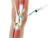
Viscosupplementation
Viscosupplementation refers to the injection of a hyaluronan preparation into the joint. Hyaluronan is a natural substance present in the joint fluid that assists in lubrication. It allows smooth movement of the cartilage covered articulating surfaces of the joint.
Glucosamine-Chondroitin Sulfate
Glucosamine and Chondroitin Sulfate are the most common nutritional/diet supplements used in the treatment of arthritis pain and inflammation. Patients who suffer from osteoarthritis may benefit from taking these supplements. Osteoarthritis, also called degenerative joint disease is the most common form of arthritis. It occurs most often in older people.
Surgical Treatment
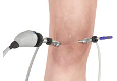
Knee Arthroscopy
Knee Arthroscopy is a common surgical procedure performed using an arthroscope, a viewing instrument, to look into the knee joint to diagnose or treat a knee problem. It is a relatively safe procedure and a majority of the patients are discharged from the hospital on the same day of surgery.
LCL Reconstruction
Lateral collateral ligament (LCL) is a thin set of tissues present on the outer side of the knee, connecting the thighbone (femur) to the fibula (side bone of lower leg). It provides stability as well as limits the sidewise rotation of the knee. Tear or injury of the LCL may cause instability of the knee that can be either reconstructed or repaired to regain the strength and movement of the knee.
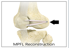
MPFL Reconstruction
Patella (knee cap) is a protective bone attached to the quadriceps muscles of the thigh by quadriceps tendon. Patella attaches with the femur bone and forms a patellofemoral joint. Patella is protected by a ligament which secures the kneecap from gliding out and is called as medial patellofemoral ligament (MPFL).
Tibial Tubercle Osteotomy
Tibial tubercle osteotomy is a surgical procedure which is performed along with other procedures to treat patellar instability, patellofemoral pain, and osteoarthritis. Tibial tubercle transfer technique involves realignment of the tibial tubercle (a bump in the front of the shinbone) such that the kneecap (patella) traverses in the center of the femoral groove.
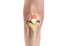
Osteochondritis Dissecans/OCD Repair
The knee, mostly the femoral condyles are most commonly affected. The two femoral condyles make up for the rounded end of femur (thighbone). Each knee has two femoral condyles, the medial femoral condyle on the inside of the knee and the lateral femoral condyle on the outside of the knee. Osteochondritis dissecans occurs within the lateral aspect of the medial femoral condyle. The condition can also occur in other joints, including your elbows, ankles, shoulders and hips.
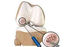
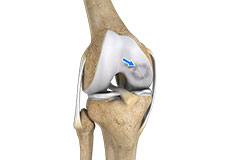
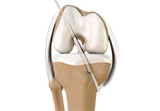
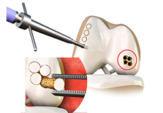
Knee Microfracture
Knee microfracture surgery is a technique to treat chondral and cartilage injuries involves stimulating the formation of new articular cartilage by drilling numerous tiny holes in the bone underneath the damaged cartilage. This results in the formation of blood clots within the damaged cartilage, which stimulates the growth of new cartilage known as fibrocartilage. Although, the fibrocartilage formed is different from the normal hyaline cartilage, it can provide significant improvement in the symptoms.
Bone Marrow Aspirate/BMA implantation
Bone marrow contains a high number of stem cells or precursor cells that have the potential to replicate and transform into cells of any tissue. It may be aspirated and concentrated to form a biologic therapy agent that is particularly useful for the healing of bone, cartilage, and tendon. It also contains growth factors, progenitor cells, platelets and white blood cells which aid the healing process.
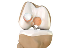
Cartilage Repair & Transplants
Cartilage replacement is a surgical procedure performed to replace the worn-out cartilage with new cartilage. It is usually performed to treat patients with small areas of cartilage damage usually caused by sports or traumatic injuries.
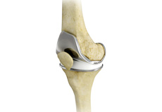
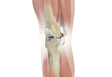
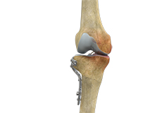
Total Knee Replacement
The knee is made up of the femur (thighbone), the tibia (shinbone), and patella (kneecap). The meniscus, the soft cartilage between the femur and tibia, serves as a cushion and helps absorb shock during motion. Arthritis (inflammation of the joints), injury, or other diseases of the joint can damage this protective layer of cartilage, causing extreme pain and difficulty in performing daily activities.
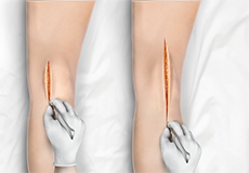
Minimally Invasive Knee Replacement
Total knee replacement is a very successful surgical treatment for knee arthritis. Over the years, minimally invasive knee replacement surgical techniques have been developed to lessen tissue trauma and improve patient outcomes. This minimally invasive approach involves much smaller incisions than the usual 10-12 inch incisions used in the traditional knee replacement and spares the quadriceps muscle and tendon, which control bending of the knee, from being cut to access the knee joint.
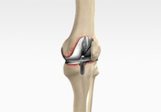
Revision Knee Replacement
Revision knee replacement surgery involves replacing part or all your previous knee prosthesis with a new prosthesis. Although total knee replacement surgery is successful, sometimes the procedure can fail due to various reasons and require a second revision surgery.
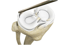
Partial Meniscectomy
Partial meniscectomy is a surgical procedure to remove the torn portion of the meniscus from the knee joint. Meniscus is the C-shaped cartilage located in the knee that lubricates the knee joint, acts as a shock-absorber, and controls the flexion and extension of the joint.
Meniscus Repair Surgery
A meniscus tear is the commonest knee injury in athletes, especially those involved in contact sports. A sudden bend or twist in your knee can cause the meniscus to tear. This is a traumatic meniscal tear. Elderly people are more prone to degenerative meniscal tears as the cartilage wears out and weakens with age.
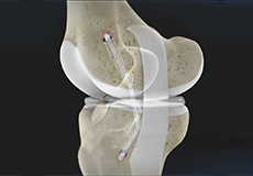
ACL Reconstruction
The anterior cruciate ligament is one of the major stabilizing ligaments in the knee. It is a strong rope- like structure located in the center of the knee running from the femur to the tibia. When this ligament tears unfortunately, it does not heal and often leads to the feeling of instability in the knee.
MCL Reconstruction
Medial collateral ligament (MCL) is one of four major ligaments of the knee that connects the femur (thigh bone) to the tibia (shin bone) and is present on the inside of the knee joint. This ligament helps stabilize the knee.
PCL Reconstruction
Posterior cruciate ligament (PCL), one of four major ligaments of the knee is situated at the back of the knee. It connects the thighbone (femur) to the shinbone (tibia). The PCL limits the backward motion of the shinbone.
Osteochondral Autograft Transfer System (OATS)
Osteochondral autograft transfer system is one of the two types of cartilage transfer procedures and the other procedure is “Mosaicplasty”. Cartilage transfer procedures involve moving healthy cartilage from a non-weight-bearing area of the knee to a damaged area of the cartilage in the knee. In mosaicplasty, plugs of cartilage and bone are taken from a healthy cartilage area and moved to replace the damaged cartilage of the knee.